Blessed and favoured: 23-hour surgery that saved miracle twins
What you need to know:
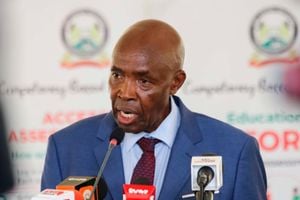
- Dr Christopher Musau is a specialist neurosurgeon with a wealth of experience spanning two decades.
- Initial preparations of the life changing operation began at 6.30am on Tuesday with the girls being wheeled into the hospital’s main theatre.
- Prior to this, the little girls had undergone a series of investigative tests for the medics to know what they were sharing.
- Blessing and Favour were conjoined at the lower back and buttock area, called pygopagus.
- For the next two-and-a-half hours, Blessing and Favour were prepped for surgery until they drifted into sleep under anesthesia.
Dr Christopher Musau and his team knew it was their turn to go into the theatre when they got the signal from the anaesthetists that conjoined twins Blessing and Favour Mukiri were sound asleep and their vital signs were stable.
Twenty-three hours later, they had completed one of the most significant and delicate operations in Sub-Saharan Africa.
Donned in short-sleeved cotton suit before entering the surgical suite, he led a team of about 27 specialists — nine paediatric surgeons, eight neurosurgeons and 10 plastic surgeons.
The neurosurgeon began his day at 4am by doing a series of exercises and a morning prayer at the All Saints Cathedral before driving up Upperhill road to Kenyatta National hospital’s main theatre.
“Whenever I have a big surgery to handle, I love going for some exercises just to put my body in the right state to withstand the long hours,” he says.
Dr Musau is a specialist neurosurgeon with a wealth of experience spanning two decades.
He has pioneered the field. In his tenure as a specialist neurosurgeon, the 59-year-old doctor who has a fetish for bowties has been able to successfully carry out intricate surgeries including one to remove a bullet lodged in the head of one-year-old Satrine Osinya, who was in 2014 shot in a terrorist attack Mombasa County.
But he is a man who is not quick to bask in the glory of the successes of surgery as he gives credit to a team of other specialists he worked with to oversee the successful 23-hour surgery on babies Blessing and Favour.
“We had planned adequately and everything went as planned,” he says.
Acknowledging that the operation was made possible by a team of neurosurgeons, paediatric and plastic surgeons, Dr Musau says that each of the 60-member team went into the operating table fully aware of the responsibility that rested on their shoulders.
“Of course there was anxiety so much so that despite the hospital providing food, most of us did not feel hungry.”
For this particular procedure, he went in knowing that it would be performed in three conclusive phases, lasting 23-hours.
Prior to this, the little girls had undergone a series of investigative tests for the medics to know what they were sharing. Soon after turning two years old, they had their first reconstructive surgery where paediatric surgeons inserted tissue expanders to stretch their skin to sure there was enough to cover their new separated bodies.
Tuesday was the second and main stage of the separation process. Although he had never separated twins conjoined at the sacrum (pygopagus surgery), a triangular bone in the lower back situated between the two hipbones of the pelvis, he terms this particular one as special. And he has learned a lot from it.
LOWER BACK
Blessing and Favour were conjoined at the lower back (sacrum) and buttock area, called pygopagus.
These twins were back-to-back facing opposite directions. In most instances, such twin share part of the lower digestive tract (large intestine, rectum and anus) and parts of the skeleton, nervous system and genitals. About 18 per cent of all conjoined twins are in this group.
For the two girls, they shared a rectum, pelvic organs, spinal cord at the lower back, spinal nerves that powered the movement of their legs and genitalia.
“We knew where we were going to start from and what steps would follow. The game plan was to start from the front, so as to free all the pelvic organs from the bone of the spine before proceeding to the back side,” he explains.
“A good surgeon will tackle any surgery as long as they know the anatomy well.”
But he had a point of concern: this surgery would determine whether after separation the girls would breathe again or not. So he says, everything had to be done with utmost precision.
“We were extra careful with the nerves because if we gave the wrong nerve or cut more than required, we would create a deficit that would likely cause paralysis. We did not want that to happen,” says Dr Musau.
Initial preparations of the life changing operation began at 6.30am on Tuesday with the girls being wheeled into the hospital’s main theatre where a team of two consultant anaesthetists (flanked by a backup team of another six support staff undergoing training equally divide to each child) were in waiting.
Besides setting up the machines to receive the babies, they were tasked with putting them to sleep and sterilising them as well as monitoring their vital signs, including if they were breathing normally.
For the next two-and-a-half hours, Blessing and Favour were prepped for surgery until they drifted into sleep under anesthesia.
At 9am Dr Musau and Dr Fred Kambuni—the hospital’s chief paediatric surgeon and a lead surgeon—accompanied by a team of neurosurgeons, plastic and paediatric surgeons walked into the main theatre, situated on the first floor of the hospital.
The medics were armed with a three-dimensional replica of the girls’ skeletal image showing the sacral bone and organs to be pulled apart, revealing exactly how they are fused together. The next several hours were a whir of activity.
“It is from the 3D image that we knew exactly what to pull apart and to what precision it needed to be,” Musau says.
Often, paediatric surgeon Dr Kambuni says, surgeries to separate conjoined twins may range from very easy to very hard, depending on the point of attachment and the internal parts that are shared.
“This particular one needed a multi-disciplinary approach and good coordination for everything was working out seamlessly. For instance, when we cut at the skin level, plastic surgeons had to be involved to mobilise adequate skin that would then cover up the wound left after the surgery,” he explains.
THREE PHASES
The surgery would then be done in three phases: phase one which began at 10am and took six hours entailed dissecting the pelvis which was done by paediatric surgeons assisted by neurosurgeons.
“The neurosurgeons had to take care of the nerves while identifying the rectum of both children as where they were joined. We also had to open up their birth canals and push them away from the spine,” Dr Musau says.
After opening up the pelvis, the girls were flipped at an angle of 180 degrees to enable the neurosurgeons to work on the spine by opening up the Dura matter—a membrane that covers the spinal cord— separating the nerves and the sacral bone.
“We needed to expose the spinal cord so that we could see the nerves and appropriately separate the ones that supply the lower limbs.”
At 12.30am in the night Blessing and Favour were successfully separated, allowing for the paediatric surgeons to come back and complete the process by separating their intestines.
“The intestines are usually the last to be separated because it is a dirty area which can predispose the patient to infections,” explains Dr Kambuni, who was leading a team of nine paediatric surgeons.
The second phase of the surgery which took about an hour began at 4am when plastic surgeons coming in to close the wounds.
The final phase of the surgery began at 5am where Dr Kambuni’s team created a stoma, an opening on the front of the girls’ abdomen to divert faecal matter and urine into a pouch (bag) on the outside of their bodies.
At 6am Blessing and Favour were wheeled out of the theatre into the Intensive Care Unit (ICU) to recuperate.
Generally, the doctors assert that the surgery was successful. But even so, the doctors were worried about the safety.
“If you are going to approach a particular organ, knowing how you are going to leave it after dealing with it was a real challenge,” notes Dr Kambuni, adding “We were so afraid of causing damage that was not present because unlike tissues, once damaged, nerves cannot be repaired.”
The age of the girls also posed a challenge as their organs were small, requiring the doctors to use microscopes to magnify them.


