Vet on call: Beware of this silent but stubborn disease
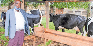
Kibira Muchai with some of his dairy cows at Riboti Farm in Elburgon, Nakuru County. The Mycobacterium avium of the subspecies paratuberculosis- abbreviated as MAP infects cattle when the animal consumes contaminated feed, water or during suckling for calves. PHOTO | JOHN NJOROGE | NATION MEDIA GROUP

What you need to know:
- Johne’s disease is both a herd and individual animal disease. It is caused by Mycobacterium avium of the subspecies paratuberculosis- abbreviated MAP.
- Generally, the Mycobacterium bacteria species cause a long-standing disease, has long incubation periods and are very difficult to treat.
- MAP infects cattle when the animal consumes contaminated feed, water or during suckling for calves. The bacteria are initially in the environment.
- Some calves may also be infected in the uterus if the mother has advanced disease where the bacteria have already entered the blood stream.
Some livestock diseases have no directly observable impact on herd production because their effects are masked by their long incubation period.
Their losses also overlap with the impacts of other production factors such as worms, some bacterial diseases and poor feeding.
The lack of suitable diagnostic facilities and public awareness also discourages farmers and animal health service providers from taking keen interest in the diseases.


I tentatively have a case of such a disease on a farm where the owner requested anonymity until I confirm the diagnosis.
Jessy, the farm’s manager, called me on phone last week to report a two-year-old heifer that had severely gone downhill in a few days.
I knew the history of the heifer as one which was generally in good health but had lagged behind its peers in growth.
I had seen the heifer a week before Jessy’s call while on a routine farm visit. Jessy had told me the heifer had had intermittent diarrhoea but at the time of my visit, the diarrhoea had stopped and the cow was eating fairly well. I told him to complete the treatment she was giving and report the progress.
When Jessy called, the heifer had just completed treatment the day before. The diarrhoea had resumed and the animal was not eating. I asked him to check all the heifer’s vital parameters and report to me soonest.
“Sorry Doc, the heifer has died even before I completed examination,” Jessy reported after 10 minutes. She further said the animal was draining out a lot of thin greenish fluid through the mouth.
This was not surprising because it had been drinking lots of water in the past 24 hours in order to replace the one that was being lost in the diarrhoea.
Since the farm is about 100km from Nairobi, it did not make sense for me to travel there to carry out post-mortem examination.
Furthermore, it was already 2pm and the curfew is on. I instructed Jessy to systematically open up the body and take photos of all the organs before and after slitting them open.
HIGHER RATES THAN PRESUMED
When I got the photos, the problem was evident in the rumen and the upper part of the large intestines. This intestinal segment is medically called the proximal colon.
Deep ulcers with dead tissue were visible in the inner walls of both organs. The proximal colon also had fresh ulcers of various sizes.
I would have liked to see the status of the glands around the intestines, called mesenteric lymph nodes, but sadly none had been captured in the photos.
The kidneys, heart, spleen and lungs showed effects of heavy bacterial infection. I formed my opinion of diagnosis as one of the neglected wasting diseases of cattle and other ruminants.
Since I have never come across the disease in my training and practice, I consulted two veterinary scientists who have research experience with the disease.
I shared the pictures and the animal’s history. Both Dr Wamalwa and Dr Omega agreed with my tentative diagnosis of Johne’s disease (JD).
They suggested I do a thorough investigation by collecting samples from adult animals in the herd and sending them to the government Veterinary Investigation Laboratory in Kericho, which Dr Wamalwa heads.
Dr Omega has carried out extensive research on the disease in Kericho County and confirmed that out of 100 cattle in the area, 16 have the disease.
Scientifically, this is called a 16 per cent prevalence rate. The national prevalence rate is yet to be determined but the Kericho one shows the disease may be there in higher rates than presumed.
The carcass had been disposed of by the time I studied the photos. The following day, I explained my findings to the farm manager and the farm owner and we agreed I would collect samples from the herd for laboratory investigation.
Johne’s disease is both a herd and individual animal disease. It is caused by Mycobacterium avium of the subspecies paratuberculosis- abbreviated MAP.
ENVIRONMENTAL CONTAMINATION
Generally, the Mycobacterium bacteria species cause a long-standing disease, has long incubation periods and are very difficult to treat.
Other species in this group include Mycobacterium bovis that causes cattle tuberculosis, Mycobacterium tuberculosis that causes human tuberculosis and Mycobacterium leprae, which causes leprosy in humans.
There is also Mycobacterium avium, the cause of tuberculosis in birds. All the mycobacteria species are very difficult or not possible to treat because they reside inside the host cells and they also have very tough protective bacterial cell wall.
MAP infects cattle when the animal consumes contaminated feed, water or during suckling for calves. The bacteria are initially in the environment.
When ingested by animals, they multiply in the intestinal cells and are shed out in faeces over many years. This increases environmental contamination in areas with infected herds.
Some calves may also be infected in the uterus if the mother has advanced disease where the bacteria have already entered the blood stream.
The bacteria mostly settle in the walls of the small intestines and the proximal colon. They are engulfed by the body’s defence cells, called macrophages, and transported to the lymph nodes, which react by producing more defence cells and chemicals called cytokines. These move to the infected intestinal walls and cause the thickened ridges and ulcers.
The thickened intestinal walls get damaged and are unable to absorb water and other nutrients. Initially, this results in intermittent diarrhoea.
Later, the diarrhoea becomes heavy and forceful, sometimes projecting far from the anus. Infected animals have very good appetite until the last stages of the disease.
However, they have progressive weight loss and mainly die in a heap of bones due to malnutrition. There is no treatment for the disease.
Infected animals in the herd are detected through laboratory testing and destroyed. Most affected animals are over two years but cases have been reported as early as six months of age.