Vet on Call: The curious incident of cow that died on Christmas Day
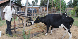
Leonard Langat feeds his dairy cows at Simotwet Village in Elburgon, Nakuru County. Careful introduction and feeding of high carbohydrate diet to livestock is essential in preventing liver abscesses. PHOTO | JOHN NJOROGE | NMG.

What you need to know:
- Liver abscesses can affect cattle of any age. The disease is common in feedlot and dairy cattle that are fed high carbohydrate diets either to fatten them or increase milk yield.
- Animals which get lactic acidosis may also, through a process that scientists are yet to figure out, cause inflammation of the hooves called laminitis.
- Most cases of liver abscess in cattle are diagnosed at slaughter or post mortem examination after dying or being sacrificed.
- Animals on high carbohydrate rations should also be given sufficient grass to provide the fibre required for rumen movement.
Veterinary services are legally classified as essential because they involve saving of animal lives, reducing suffering thus safeguarding their welfare and providing peace of mind to animal owners.
It is on this basis that I found myself on duty on Christmas Day. The year has been quite busy and I had hoped that one of the other doctors would hold the fort on the day.
Little did I recall that I was off duty on last year’s Christmas Day. The duty hammer, therefore, fell squarely on me.


The day did not disappoint. I had two dog emergency cases by 10am. I had hardly rested after attending to the cases when Kilonzo, the manager of a dairy farm in Thika called.
He informed me he had a cow with breathing difficulties and he was not sure if it was an emergency.
I requested for the full case history. Kilonzo explained that the cow was one of the two first-time mothers that had calved in October.
There had been no apparent problem with the cow but she appeared to have been losing weight since calving. The loss was not severe and Kilonzo had been watching the animal planning to report if more serious signs of illness were observed.
The cow’s milk production had dropped to two litres in the morning. Kilonzo had wanted to see whether the cow would compensate milk production in the evening before reporting suspected illness to me.
The animal had been mounted by another cow on heat that morning and it fell down. It had woken up after about 40 minutes and started having severe breathing difficulties. “Could the mounting have caused some internal injuries to the cow?” he posed.
I told Kilonzo it was unlikely the cow had been injured by the mounting. In any case, if injury was causing the raised breathing, then the cow should have been showing other signs of pain such as granting, limping and unwillingness to move.
I instructed Kilonzo to take the cow’s rectal temperature and inform. The report he made did not sound good. I was also cautious because the farm had lost about seven animals this year due to nerve damage, milk fever and an outbreak of the deadly triad of anaplasmosis (gull sickness), babesiosis (redwater) and east coast fever.
AFFECT CATTLE OF ANY AGE
I had not even completed checking and packing my drug kit when Kilonzo called with a sense of urgency, “Doc, the cow is down, kicking and frothing from both nostrils. The temperature is 36 degrees Centigrade and I think she’s dying,” he shouted inadvertently.
I told him I would be there in about 30 minutes since I expected light traffic on Thika Superhighway.
At Juja, I realised how wrong my projection was. The traffic was snaking all the way to Blue Post Hotel. I ended up taking almost two hours to arrive on the farm thanks to 12 vehicles that had stalled on the road.
By the time I arrived, the cow had been dead for 20 minutes. White froth and blood-tinged fluid was oozing from the nostrils. I kitted up and proceeded with the post-mortem examination.
The lungs were characteristically swollen, liver-like and contained a lot of bloody fluid and froth. The chest cavity was full of watery reddish fluid instead of the normal scanty straw coloured to clear fluid.
Some lung lobes were adhering to the diaphragm. There was pus in the chest cavity and the lungs. Part of the small intestines was infected with bacteria causing bleeding. The uterus had some mild bacterial infection.
Another major problem was in the liver. One of the liver lobes had a huge abscess containing thick greenish-yellow cheesy pus.
The abscess had a very thick capsule. Part of the liver and the reticulum was adhering to the diaphragm. This was most likely the epicentre of the fatal infection.
“Kilonzo, your cow died of chronic suppurative pneumonia and liver abscess,” I gave my diagnosis. I further explained to the manager that no treatment would have saved the cow. The prognosis or chance of recovery, for cattle affected with the disease is usually very poor.
Liver abscesses can affect cattle of any age. The disease is common in feedlot and dairy cattle that are fed high carbohydrate diets either to fatten them or increase milk yield.
The high carbohydrate rations cause acidity of the rumen contents, which inflames the internal surface of the rumen and causes bacteria to infect the rumen tissues.
CAREFUL FEEDING OF HIGH CARBOHYRATE DIET
Once the bacteria get into the rumen tissues, they may find their way into the liver through the blood vessels. Some of the bacteria are trapped in the liver where they multiply and form abscesses.
The bacteria then move from the liver abscess and get into the lungs through the heart. In the lungs, the bacteria cause pneumonia and formation of pus.
Animals which get lactic acidosis may also, through a process that scientists are yet to figure out, cause inflammation of the hooves called laminitis.
Bacteria may infect the inflamed feet and then spread to the rest of the body through the blood stream. The bacteria may again be trapped in the liver where they cause formation of abscesses.
Cattle with liver abscesses have poor performance in fattening and milk production. They may also have bouts of fever.
The condition is, however, very difficult to promptly and accurately diagnose in cattle but can be suspected by analysing laboratory results, case history and the clinical findings when the animal is examined.
Most cases of liver abscess in cattle are diagnosed at slaughter or post mortem examination after dying or being sacrificed.
Scientifically, we sacrifice an animal to carry out post-mortem examination to make a diagnosis for the benefit of other animals.
Many types of bacteria may cause the liver abscesses but the most common are in the groups Fusobacteria, Staphylococcus, Streptococcus, Truepelellar and Bacteroides.
Prevention of the disease in cattle is by careful feeding of high carbohydrate diet to ensure that animals are introduced to the diet slowly.
Animals on high carbohydrate rations should also be given sufficient grass to provide the fibre required for rumen movement.
After my explanations, Kilonzo sighed with relief from the knowledge that he and his team had not been negligent.