Easier, faster way to test for TB
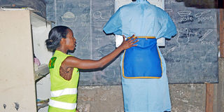
Millicent Mugo from the Ministry of Health directs a patient undergoing a chest x-ray during the launch of Kenya Tuberculosis Prevalence Survey for 2015-2016 at Mshomoroni area in Mombasa during a past event. PHOTO| FILE| NATION MEDIA GROUP

Scientists have created a glowing probe that can identify and light up single specimens of the bacteria that cause tuberculosis (TB).
In a process using bacteria that glows during tests, researchers have developed an imaging technique that can diagnose live tuberculosis samples within an hour and monitor the effectiveness of treatments.
TB, which is caused by Mycobacterium tuberculosis, is a highly contagious disease that infects the lungs, causing coughing, fever and weight loss.
In Kenya, the national TB prevalence survey in 2015 found that there are more TB cases than previously estimated. The survey found that the current practice of screening for TB symptoms and using microscopy as the only test misses many cases. According to survey findings, use of the Gene Xpert test resulted in diagnosis of 78 per cent of TB cases.
To diagnose TB, clinicians collect a sputum sample, which is cultivated in the lab for bacteria to grow to detectable levels. This requires specialised facilities.
COULD SPREAD DISEASE
The new probe and a microfluidic chip that counts TB bacteria within a sample can make the process easier. The microfluidic chip is a set of micro-channels on glass, silicon or a polymer. The micro-channels are linked, allowing scientists to mix, pump, sort or control the biochemical environment.
“Current methods can take up to two months to complete. During this time, an infected person whose status is yet to be confirmed could spread the disease to many others, even if he doesn’t know he is infected. A quicker diagnosis could curtail the infection rate,” said senior author Prof Jianghong Rao, adding that there is urgent need for quick and effective testing techniques.
The probe is effective in detecting the enzyme Blac, which breaks down the structure of many common antibiotics and fuels drug resistance. The research team designed a molecule that is activated by Blac and that also attaches to another enzyme called DprE1. When activated by Blac, the probe produces a bright green colour in an hour, allowing for rapid labelling and identification of both single and multiple TB bacteria.
Scientists tested the probe in a weakened variant of TB and found it could distinguish between live bacteria and dead bacteria, as well as between TB species and 43 other bacterial species. Live bacteria glow with a green colour, while dead or different bacteria species appear dark.
This method is cheaper and easier to carry out. It uses regular fluorescence microscopes that nearly all hospitals have and that require no specialised training. All that is required is a sample of the patient’s sputum which is then analysed using a microscope.
If the technology is approved, it can be rolled out for use worldwide. The findings were published in the journal Science Translational Medicine.